


The spinalis is one of three muscles that comprise the erector spinae (erect = upright and spinae = spine) group of muscles. The iliocostalis and longissimus are also part of this group. These muscles span the entire posterior trunk and connect the sacrum, ilium, vertebral column, and skull, running parallel to the vertebral column. The erector spinae group comprises the intermediate layer of muscle on the posterior trunk, found deep to the latissimus dorsi, trapezius, and serratus posterior muscles and superficial to the semispinalis, multifidi, and rotatores.
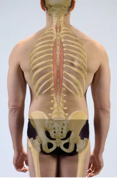
The spinalis is the most medial of the three erector spinae muscles. It extends from the spinous processes of the lower thoracic and upper lumbar vertebrae superiorly to those in the upper thoracic and lower cervical vertebrae. Its fibers run vertically, making it stronger in extension and lateral flexion than rotation compared to the longissimus and iliocostalis. In the cervical spine, the spinalis joins the semispinalis muscle of the transversospinalis group before attaching to the occiput. The semispinalis, multifidi, and rotatores muscles make up the transversospinalis group.
Functionally, the spinalis and other erector spinae muscles provide broader stabilization and movement than the deeper transversospinalis group. Together, the erector spinae and transversospinalis groups maintain upright posture of the spine against gravity. The deeper, smaller muscles of the transversospinalis group position individual or pairs of vertebrae, while the more superficial, larger muscles of the erector spinae position broad segments of the vertebral column and initiate and control movements including extension, lateral flexion, and rotation of the trunk.
Weakness in the deeper transversospinalis muscle group may cause compensation by the overlying erector spinae group. This presents as hypertonicity in the erector spinae muscles, decreased mobility in the individual joints of the vertebral column, and overall stiffness in the movements of the spine as large segments of the vertebral column move as units. Reducing overactivity in the erector spinae muscles and restoring function of the underlying transversospinalis muscles is essential to a healthy spine and functional movement.
Palpating the Spinalis
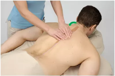
Positioning: client prone.
-
Standing at the client’s side facing their spine, locate the thoracic spinous processes with the fingertips of both hands.
-
Slide your fingertips laterally, past the lamina groove onto the erector spinae muscles.
-
Strum back and forth across the erector spinae muscles with the fingertips of both hands, locating the most medial edge formed by the spinalis.
-
Have the client gently lift their head and extend their trunk to ensure proper location.
Spinal Mobility Quick Assessment


Positioning: client in quadruped (hands and knees).
- Stand at the client’s side with an unobstructed view of their spinal curvature.
- Instruct the client to “round” their spine into an upward curve.
- Next, have the client relax, allowing their spine to “sag” into a downward curve.
- Look for individual segment motion with each movement and note any areas that remain “flat,” as segments move in groups.


