


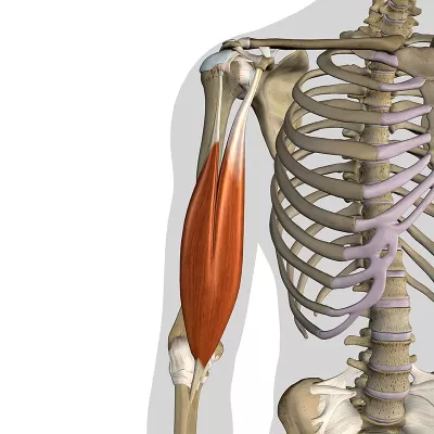
The biceps brachii muscle is one of the most well-known muscles in the human body. Ask anyone to flex their biceps, they will probably roll up their sleeves to show off their “guns.” But how well do we know the biceps story? Beyond the muscle belly and tendon, a lesser-known part of the biceps muscle may have more to do with the function of the whole arm than you would think. Allow us to reintroduce you to the lacertus fibrosus—a broad, flat fascial structure that extends across the cubital fossa and is credited with some fairly big capacities, including the ability to improve muscular efficiency, enhance grip strength, and assist with neuromuscular coordination.

The lacertus fibrosus, also known as the biceps aponeurosis, is one of the most recognized myofascial expansions in the body. While the biceps brachii tendon (top left) roots deep into its bony insertion at the radial tuberosity, the lacertus fibrosus, visible here as a sweep of white collagen “threads” (center to bottom right), crosses more superficially, fanning out and merging with the fascial sleeve of the forearm, the antebrachial fascia. Because of this three-dimensional organization of insertion, flexing the biceps relays force to the radial bone and simultaneously tensions the antebrachial fascia, increasing biomechanical efficiency, and supporting the body’s ability to perform complex movements with fluidity. Image courtesy AnatomySCAPES.
The Biceps Brachii “Origin” Story
The biceps brachii muscle is a two-headed fusiform muscle that assists with shoulder flexion and contributes to shoulder stability but is best known for its actions of elbow flexion and supination, as in the action of showing off our “guns.” The two heads find their origin on the scapula, while the single distal tendon inserts at the radial tuberosity. But, if you look carefully in your anatomy textbook, there’s often a second insertion point listed: the deep fascia of the forearm.
Here’s where things get interesting. The biceps brachii tendon inserts deep into the radius bone, but the biceps brachii fascia has its own much broader destination. Fanning out from the tendon, it heads toward the surface, inserting into the highly innervated antebrachial fascia—the membranous deep fascia “sleeve” of the forearm. During contraction, the biceps muscle pulls on the bone while simultaneously stretching the enveloping antebrachial fascia.
This illustration demonstrates the classic understanding of how muscle becomes tendon in fusiform muscles. As the muscle approaches its origin or insertion, the red contractile muscle fibers gradually fade away while the muscle's connective tissue framework—epimysium (surrounding the entire muscle), perimysium (wrapping muscle bundles), and endomysium (encasing individual fibers)—converges and densifies to form the white, collagen-rich tendon. This represents the simplified "muscle contracts-pulls tendon-moves bone" model. What's missing is the significant role of myofascial expansions. Extending beyond the primary tendon, myofascial expansions insert into deep fascia (not bone), creating broad fascial connections and coordinating movement across multiple body segments.
Getty Images.
Put another way, imagine you have a travel bag for your massage table that allows the handles of your table to pop through the bag. In addition to your table handles, the bag itself has handles. By using both sets of handles simultaneously when you go to pick up your massage table and carry it to your car (biceps contraction), you apply force to both the table (radial bone) and the travel case (antebrachial fascia) via their handles (biceps tendon and lacertus fibrosus, respectively). This kind of fascial connection is called a myofascial expansion—a specific connection that exists between either skeletal muscles or tendons and aponeurotic deep fascia. Myofascial expansions go far beyond simple muscle-tendon-bone connections, and many, if not most, of our muscles have them. The biceps brachii myofascial expansion is so obvious that anatomists have even given it a name: lacertus fibrosus.
Lacertus Fibrosus’s Hidden Powers
The lacertus fibrosus resembles the curl of a lizard’s tail (lacertus comes from the Latin word for lizard) and is easily seen when the skin and subcutis are removed, yet scientists are still exploring its functions.
Looking at whether the lacertus fibrosus is significant biomechanically, in a cadaveric study, one group of researchers measured what happens when the lacertus fibrosus is removed.1 The effect on muscular efficiency was dramatic:
- 28 percent loss in flexion efficiency
- 50 percent loss in supination efficiency
The lacertus fibrosus appears to increase the biceps lever arm at the elbow. The more-extended lever arm saves energy and improves muscle efficiency by creating the same movement with less effort.
But the benefits of the lacertus fibrosus extend beyond simple mechanical advantage. Scientific literature describes it as a “strength strap” that creates functional synergy between the biceps and forearm flexors during grip activities. It also helps elbow flexion and forearm supination work together with what researchers call “rhythmicity”—a coordinated, flowing relationship between movements. Findings from fascia research may further explain this idea of functional synergy and rhythmicity.
A New “Origin” Story for Muscles
We might have to rethink some of our simpler models of how the body moves based on current muscle force transmission studies. It’s not just a story of muscle pulling on bones via the tendons. Studies reveal that up to 37 percent of the force generated by muscle contraction doesn’t go to the bones at all. Instead, it’s transmitted to adjacent connective tissue structures.2 In myofascial expansions, these connections work in two primary ways: fascia as insertion and fascia as origin.
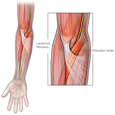
The lacertus fibrosus is one of the most well-documented myofascial expansions in the human body. It is so prominent that it appears in anatomical illustrations even when other fascial structures are omitted. This broad, flat fascial structure extends from the biceps brachii across the cubital fossa to merge with the highly innervated fascial continuity of the forearm, the antebrachial fascia. This myofascial expansion exemplifies how fascial structures can serve both biomechanical and sensory roles, demonstrating the sophisticated organization of the body’s connective tissue networks.
Image courtesy Science Direct.
Fascia as Insertion
Instead of inserting into bone, the muscular fascia expands beyond the tendon and joins with the aponeurotic deep fascia of the next region. The lacertus fibrosus is a perfect example of this in the upper limb—instead of inserting into the radius bone along with the biceps tendon, it expands beyond the tendon like a membranous sail and merges with the antebrachial fascia of the forearm.
Fascia as Origin
Rather than originating from bone, the muscle fibers are firmly anchored into aponeurotic deep fascia. In the forearm, this fascia-as-origin organization is found at the proximal end of the flexors and extensors near the elbow, where they root directly into the inside of their enveloping antebrachial fascia. The connection changes toward the distal end, where the muscles and their long tendons are free to glide beneath the antebrachial fascia.
The lacertus fibrosus tensions the antebrachial fascia during biceps contraction, creating reciprocal feedback that helps coordinate movement.
Because fascia is innervated with sensory nerve endings that perceive stretch, myofascial expansions like the lacertus fibrosus enable distal muscle groups to be informed about proximal muscle contraction states during movement. The lacertus fibrosus tensions the antebrachial fascia during biceps contraction, creating reciprocal feedback that helps coordinate movement. The entire system functions as an integrated unit, rather than isolated parts.
But the lacertus fibrosus isn’t the only place we find this kind of organization. A groundbreaking study provided the first comprehensive evidence that muscles create systematic myofascial continuity throughout the upper limb.3 The study demonstrated that the fascial insertions were consistently present across all specimens and exhibited constant anatomical patterns of myofascial expansions:
- Pectoralis major brachial fascia (two pathways)
- Latissimus dorsi posterior brachial fascia
- Triceps posterior antebrachial fascia
- Biceps anterior antebrachial fascia
- Palmaris longus palmar and thenar fascia
- Extensor carpi ulnaris hypothenar fascia (in 85 percent of cases)
Myofascial expansions like these create three-dimensional functional architecture that changes how we understand movement, from isolated muscle actions to integrated fascial networks coordinating function across the body.
Why We Care
Myofascial expansions like the lacertus fibrosus are more than just helpful second insertion points; they serve as essential elements for our entire neuromuscular coordination system. Understanding the complete anatomy of myofascial expansions like this one can help us assess and address muscles beyond their borders into their fascial continuities, ultimately leading to better outcomes for our clients, from shoulder to fingertip and head to toe!
Notes
1. Olivier Snoeck et al., “The Biomechanical Role of the Lacertus Fibrosus of the Biceps Brachii Muscle,” Surgical and Radiologic Anatomy 43, no. 10 (October 2021): 1587–94, https://proquest.com/docview/2574930908.
2. Mark J. C. Smeulders and Michiel Kreulen, “Myofascial Force Transmission and Tendon Transfer for Patients Suffering from Spastic Paresis: A Review and Some New Observations,” Journal of Electromyography and Kinesiology 17, no. 6 (December 2007): 644–56, https://doi.org/10.1016/j.jelekin.2007.02.002; Snoeck et al., “The Biomechanical Role of the Lacertus Fibrosus of the Biceps Brachii Muscle.”
3. Carla Stecco et al., “Tendinous Muscular Insertions onto the Deep Fascia of the Upper Limb. First Part: Anatomical Study,” Morphologie 91, no. 292 (March 2007): 29–37; Carla Stecco et al., “Anatomy of the Deep Fascia of the Upper Limb. Second Part: Study of Innervation,” Morphologie 91, no. 292 (March 2007): 38–43, https://doi.org/10.1016/j.morpho.2007.05.001.
Resources
Stecco, C. “Fascial Anatomy” in Fascia, Function, and Medical Applications. CRC Press, 2021.
Szewczyk, B. et al. “Anatomical Variations of the Biceps Brachii Insertion: A Proposal for a New Classification.” Folia Morphologica 82, no. 2 (2023): 359–67. https://doi.org/10.5603/fm.a2022.0022.
A Deeper Dive—Two Types of Deep Fascia
Epimysial Fascia
Thin, membranous deep fascia that envelops and intrapenetrates individual muscles, giving them form and shape, including epimysium, perimysium, and endomysium.
Aponeurotic Fascia
Thicker fibrous deep fascia that surrounds or connects groups of muscles. On the extremities, it envelops the entirety of each of the limbs as a fibrous sleeve, forming compartments that connect to the bones. On the trunk, it exists as dense, layered hubs on the abdomen and low back and is formed by multiple flat tendons.
Upper Extremity Aponeurotic Fascia
Brachial Fascia: Aponeurotic fascia of the arm (above the elbow)
Antebrachial Fascia: Aponeurotic fascia of the forearm (below the elbow)


