


Headaches are complex, with many potential causes. While massage may have indirect benefits for any type of headache, tension headaches and cervicogenic headaches have been shown to directly benefit from manual therapies.1 Massage protocols addressing tension headaches tend to focus on the muscles of the head, neck, and jaw, as well as the joints of the cervical spine.2 But what about the rest of the body? Could there be other contributing factors creating and perpetuating chronic neck tension?
For headaches, we can start by assessing the relationship of the head to the cervical spine. Because many of us sit staring at computers or phones more than our bodies would like, forward-head posture is the most common presentation, with compression at the base of the skull and tension in the sternocleidomastoid, trapezius, and deeper neck muscles. This is where standard headache protocols come in; it makes sense to address the area locally. But is it possible that even more distal patterns are perpetuating the imbalance? Let’s first explore the concept of posture.
Posture Involves More Than the Head and Neck
Many discuss and assess posture as a static destination in which all body parts are in their right place with proper alignment. But the body is a dynamic system, constantly responding to external and internal forces, like gravity and the movement of the breath cycle. And the “alignment” of the body only matters to the extent to which it allows for efficient and healthy breathing and movement.
Perhaps a better approach to posture would be to observe it as a dynamic series of relationships. Every part of the body is in a structural and functional relationship with every other part of the body. Our work is to facilitate balanced, functional relationships in which force is distributed efficiently with minimal tension and effort. One concept for depicting this is the tensegrity model—a system containing tensional elements (like our muscles, tendons, ligaments, and fasciae) and solid struts (like our bones). When there is minimal tension distributed evenly throughout the system, a balanced, lightweight, dynamic structure emerges. If there is increased tension in one part, the effects are felt throughout the system as strain or excess compression. This holds true in the body: A restriction in one area can translate through the rest of the body, producing compression, lack of function, or pain in another area.
While the neck is of course relevant for headaches, the neck and head do not live in a vacuum.
One of our great assets as massage therapists is that we get to work with the body as a whole. While the neck is of course relevant for headaches, the neck and head do not live in a vacuum—their relationship is dependent on all the other relationships in the body. There are many relevant factors we could consider beyond the neck muscles when it comes to headaches, such as restriction in the first rib and thoracic spine, tension in the stomach, or tight adductors. But probably one of the least discussed is the role of the anterior and lateral compartments of the lower leg. We consistently observe in clinical practice and trainings that addressing lower-leg myofascia can dramatically decrease neck tension and improve neck range of motion. Let’s consider some possible mechanisms.
Lower-Leg Fascia That Can Impact the Rest
The crural fascia (the fascia encasing the lower leg) is quite dense in response to the circumferential force it manages—when you step on your foot, weight doesn’t only go down, it goes out, and structures like the interosseous membrane and crural fascia are fortified to manage that force.3 Some research suggests that the crural fascia can become even more dense in response to pathological conditions such as ankle sprains or diabetes.4 Additionally, the muscles of these compartments are often underused. Most people walk only on flat, hard surfaces (i.e., sidewalks or floors) while wearing restrictive footwear. Our ankles, feet, and associated muscles are designed to adapt to uneven surfaces and absorb and generate significant forces. That means they are made to move, which improves circulation and tissue hydration, slide, and function. They benefit from dynamic training, and without it, the muscles and fasciae become rigid, stiff, and tight.

This fascial stiffness in the lower-leg compartments can reduce force absorption during walking or running, which translates up the kinetic chain. Force will have to be absorbed somewhere, reverberating in the knee, pelvis, lumbar spine, and potentially cervical spine and skull. This reverberation increases stress in the neck, head, and jaw, potentially resulting in greater tension in supporting muscles. Imagine wearing full-body footed pajamas that are too short and inflexible in the leg and foot. If you tried to zip it up, you would feel strain and compression in the shoulders and neck.
There are also myofascial continuities from the leg muscles that impact the head and neck posture and mobility. The tibialis anterior fascia is in direct continuity with the iliotibial (IT) band and works in conjunction with the tensor fasciae latae and the gluteus maximus to manage the tension of the IT band. This line is so clear to me in dissection and function that I like to teach it as the “IM band” or the “iliometatarsal band.” Excess tension in the tibialis anterior translates into the hip and can cause an array of functional and structural responses: an internally rotated tibia, dropped arch, externally rotated hip, and often a combination thereof. Either way, this translates into compensations in the upper body, such as thoracic rigidity and kyphosis, which leads to forward-head posture and neck tension.
The fibularis muscles in the lateral compartment work with the tibialis anterior and posterior to manage the foot’s adaptability, force absorption, and force generation. Tension here affects the foot’s ability to transform easily between foundation mode (for force absorption) and lever mode (for force generation). The fibularis longus relates into the IT-band pattern described previously, as well as into the biceps femoris. Via this lateral hamstring continuity (in the lab, we call it the “hamfoot”), tension in the lateral compartment can drive eversion, pronation, and lateral hip rotation, pushing the pelvis forward and leading to a posterior tilt in the rib cage, rounding of the upper thoracic spine, and compression at the base of the skull. This creates a similar result as the anterior compartment imbalance, just a slightly different pathway for getting there.
And that’s the beauty of the fascial body: There are infinite paths we could follow to connect the lower leg to the neck and head. Tom Myers has a few others in Anatomy Trains that offer slight variations on what I’ve suggested here but end with a similar result (see the lateral line and the deep front line).5 The more you trace different pathways, the more obvious it becomes (all roads lead to Rome after all). Any way you slice it, the lower leg impacts the neck and head.
Let’s pause. This is starting to sound like a lot of jargon. I don’t want you to have only technical-sounding information. The goal is for you to understand and observe variations of these patterns in clients. In our trainings, we explore through embodiment, primarily using the Franklin Method, which means trying these patterns on and experiencing them yourself. So don’t take my word for it, play around; grab your favorite anatomy atlas or app, and pull up the lower-leg muscles.
Explore Your Own Lower-Leg Fascia
Touch and trace the muscles in your body—the tibialis anterior, extensor digitorum longus, and fibularis longus and brevis. Starting at their attachments, follow the continuities described above into the IT band and biceps femoris. You could explore these lines through different superficial and deep pathways all the way to the neck and head, though their impact on pelvic orientation and movement is significant enough to affect the neck.
Now, stand up and sense what happens throughout the rest of your body when you tension certain aspects of the line—the anterior leg and lateral leg. Play with variations and different connections. Observe how the compensatory patterns cycle all the way from the foot to the head and back again. Change one thing in the foot, and you’ll feel it in the head. Change the head position, and it ripples down through the foot. Keep in mind that, while there are common patterns that can present, every body expresses the story of its unique movement history, and the fasciae and muscles reflect those unique patterns. Reflect on your experience and name what you notice in your body. As you get better at this, you also get better at assessing and addressing dynamics in your clients’ bodies.
Change one thing in the foot and you’ll feel it in the head. change the head position and it ripples down through the foot.
In fact, this embodiment approach is a powerful assessment tool; try on your client’s posture. Don’t tell them you’re assessing posture, simply observe them as you stand and chat. Starting at the feet, adjust your body to mirror theirs and notice where you feel tension, compression, or strain. This gives you an idea of where the tension is and where they feel pain or compensation.
Seeing Results in Action
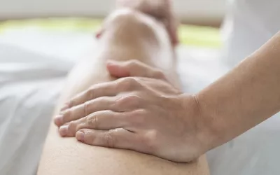
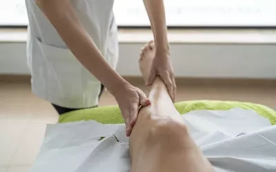
The treatment approach we have found most clinically effective is fascial therapy in the style of structural integration—slow, intentional work focused on the fascial containers rather than gliding strokes focused on superficial layers or muscles. Anecdotally, in a hybrid in-person/online foot and lower-leg training we were teaching, we hadn’t yet mentioned the neck connection. One of the online students was working with her practice partner, and after receiving the work focused solely on the anterior and lateral compartments, her partner spontaneously reported significantly reduced neck tension and pain. This happens all the time in practice, but I share this anecdote because it was a perfect example of how, even without the intention or awareness to address the neck issues, the lower-leg work had that effect.
Hopefully, the potential structural and functional mechanisms I’ve proposed for the relationship between the anterolateral leg and neck tension and posture have piqued your willingness to explore far beyond the neck when treating headaches. As an educator, I am always cautious to say, “My story about it is not the story.” The body is incredibly complex, and we do not yet have anything close to the full picture!
Even as I wrap up this article, I can think of numerous avenues we could take to tell an equally compelling story, such as meridian pathways or the nervous system. Regardless of which explanation you choose, it should not impede or diminish the treatment opportunity; addressing lower-leg myofascia can reduce neck tension, improve neck and head posture, increase mobility, and thus reduce headaches. As always, don’t take my word for it, give it a try in practice. I’d love to hear what you find.
Notes
1. C. Lozano López et al., “Efficacy of Manual Therapy in the Treatment of Tension-Type Headaches. A Systematic Review from 2000–2013,” Neurología 31, no. 6 (July-August 2016): 357–69, https://doi.org/10.1016/j.nrl.2014.01.002; Danilo Kamonseki et al., “Effectiveness of Manual Therapy in Patients with Tension-Type Headache. A Systematic Review and Meta-Analysis,” Disability and Rehabilitation 44, no. 10 (May 2022): 1780–9, https://pubmed.ncbi.nlm.nih.gov/32924640.
2. Steen Berggreen, Edith Wiik, and Hans Lund, “Treatment of Myofascial Trigger Points in Female Patients with Chronic Tension-Type Headache—A Randomized Controlled Trial,” Advances in Physiotherapy 14, no. 1 (February 2012): 10–7, https://doi.org/10.3109/14038196.2011.647333; Albert F. Moraska et al., “Myofascial Trigger Point-Focused Head and Neck Massage for Recurrent Tension-Type Headache: A Randomized, Placebo-Controlled Clinical Trial,” The Clinical Journal of Pain 31, no. 2 (February 2015): 159–68, http://doi.org/10.1097/AJP.0000000000000091.
3. Carla Stecco et al., “Mechanics of Crural Fascia: From Anatomy to Constitutive Modelling,” Surgical and Radiologic Anatomy 31, no. 7 (August 2009): 523–9, https://doi.org/10.1007/s00276-009-0474-2.
4. Carmelo Pirri et al., “Ultrasound Imaging of Crural Fascia and Epimysial Fascia Thicknesses in Basketball Players with Previous Ankle Sprains Versus Healthy Subjects,” Diagnostics 11, no. 2 (January 2021): 177, https://doi.org/10.3390/diagnostics11020177; Carmelo Pirri et al., “Crural and Plantar Fasciae Changes in Chronic Charcot Diabetic Foot: A Cross-Sectional Ultrasound Imaging Study—An Evidence of Fascial Continuity,” Journal of Clinical Medicine 12, no. 14 (July 2023): 4664, https://doi.org/10.3390/jcm12144664.
5. Tom Myers, Anatomy Trains: Myofascial Meridians for Manual Therapists and Movement Professionals, 4th ed. (Elsevier, 2021).


